baby chest x ray technique
Up to 10 cash back The reject rate for the standard mobile chest X-ray technique is typically 67 approximately 18000 performed annually. Use only if necessary.

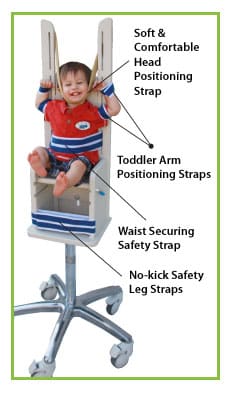
Pdf Radiation Protection In Pediatric Radiography Introducing Some Immobilization And Protection Equipment Semantic Scholar
Inspiration Penetration Rotation is part of the Lecturio course Radiology WATCH the complete course on httplect.

. Full legfull spine imaging is performed at 180 cm using CR. It is chiefly used in the pediatric population. Indication Undertaken to demonstrate small pleural effusions or for the inv.
Lie on an X-ray table on your side with your ear glued to. Whilst many of the radiological appearances are relatively non-specific integration of the clinical features with the X-ray. A chest X-ray is a safe and painless test that uses a small amount of radiation to take a picture of a persons chest.
This video Chest X-Ray Techniques. Chest or Thoracic Structures CT Scan CT imaging of the chest presents unique challenges because. Indications This view is preferred in infant and neonate imaging whilst AP erect and PA erect views are ideal for chi.
Central Ray The central ray is an imaginary -ray that comes rightx down the center of the entire -ray beam. The American Dental Association ADA recommends that kids and teens get bitewing X-rays every six to 12 months if they have cavities. Chest lateral 180 cm.
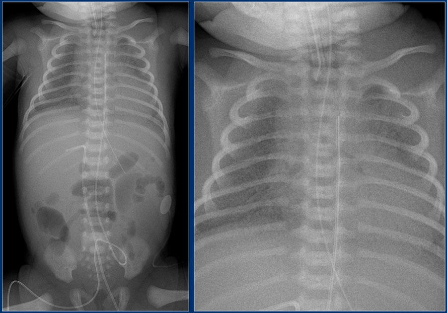
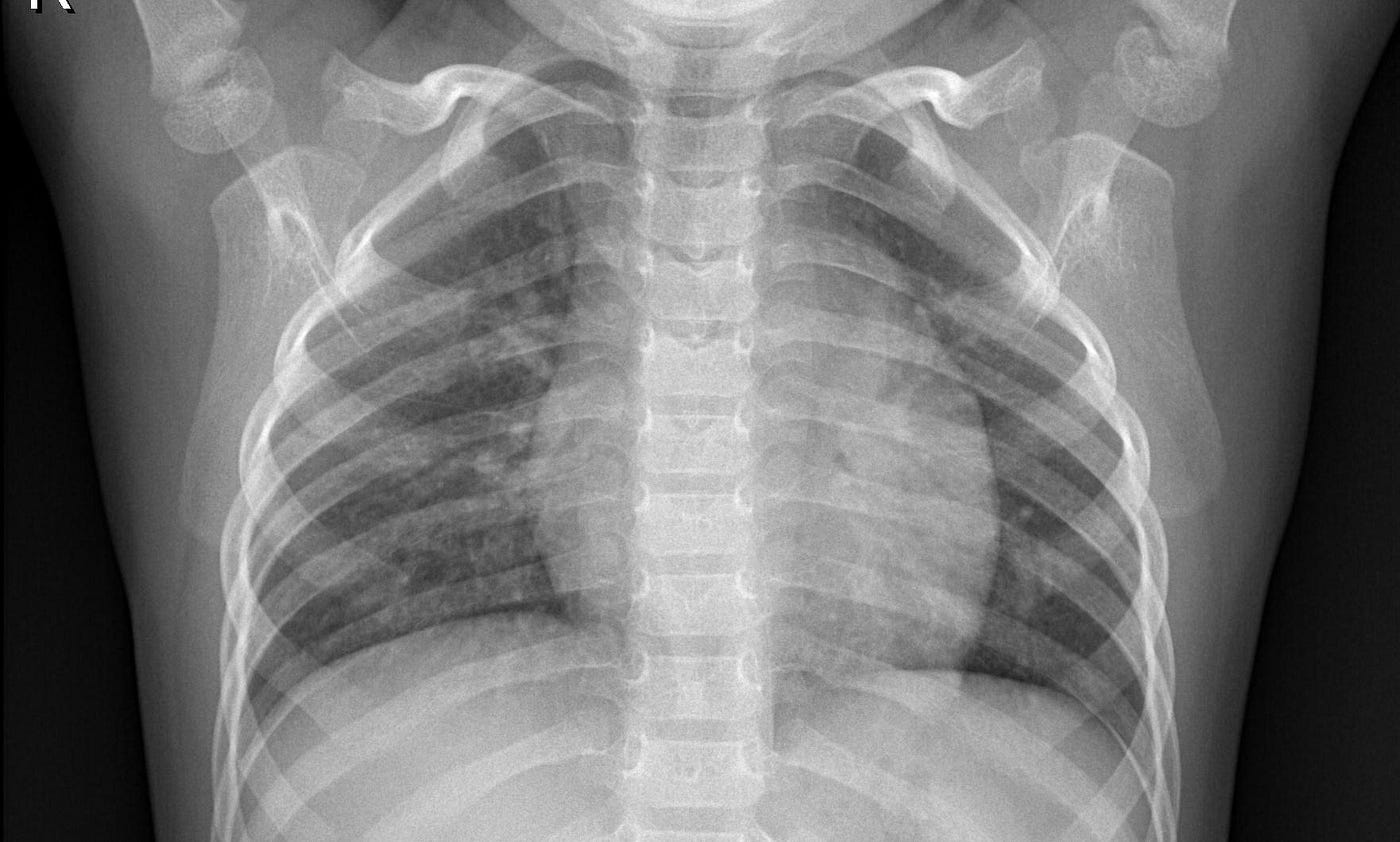
The initial view is from the front and the second is a side view. Medical X-ray imaging has led to improvements in the diagnosis and treatment of numerous medical conditions in pediatric patients. The chest X-ray technique in young children involves two views.
Interestingly this improved and dropped to 8 in the second month possibly as radiographers became more competent. GE AMX4 portable x-ray system Fuji CR imaging plates and reader Tracked AP chest and abdomen for patients 0-3 months in the NICU and PICU at Hadassah Medical Organization Image quality assessment and dose estimation for high and low kVp image sets. However all children are modest to some degree about having their genitals or backsides exposed after ages 4 to 5.
In pediatric imaging the anteroposterior supine chest x-ray is beneficial for imaging unconscious or uncooperative patients. We use the central ray CR tox point the -ray beam where we want it to go. X-ray examination of KUB on infants - AP Abdominal X-ray.
This technique represents the expansion in two dimensions only. Most neonatal chest X-rays are AP films unless the baby is made to lie prone Lucency of soft tissue shadow - darker the soft tissue more. Semianthropomorphic phantoms of the head abdomen and pelvis were placed adjacent to a chest phantom to mimic the habitus of a 5-year.
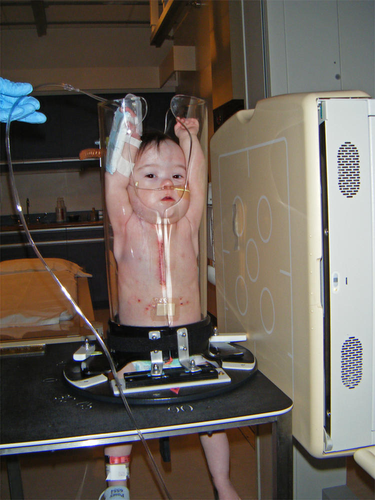
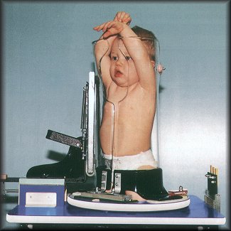
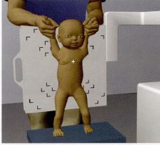
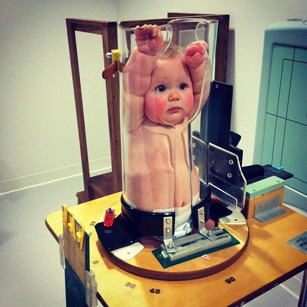
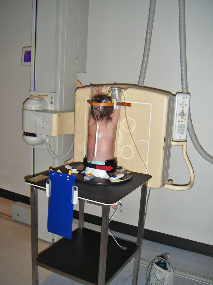
If the infant will require intubation for greater than 10- 14 days consider the use of a palate plate to. In contrast most 12-year-old males have little modesty about their chests. In young children the patient lies on the table and the hands are held above the head.
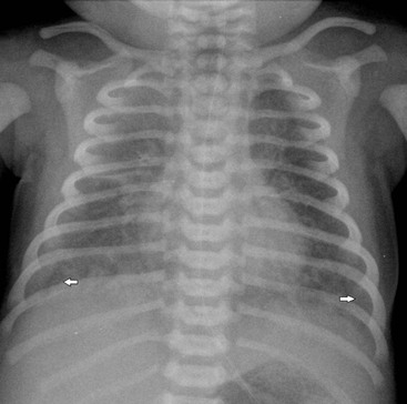
During the examination an X-ray machine sends a beam of radiation through the chest and an image is recorded on special film or a computer. For example a standard X-ray of the chest provides about the same amount of radiation that you would normally get from background environmental radiation in 2 to 3 days. A chest x-ray is frequently performed in infants with LRTI caused by RSV.
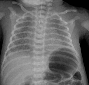
Schematically read and describe a neonatal chest X-ray 2. But newborns unlike older infants and children have. Chest lateral supine 110 cm.
9025a a Use automatic exposure control 500 speed for chestabdomen else 400 speed at specified kVp when practical. 7725a a Use automatic exposure control 500 speed for chestabdomen else 400 speed at specified kVp when practical. Consideration will be given to neonatal anatomy and development the importance of good preparation radiographic technique and positioning exposure factors radiation protection and optimal imaging outcomes.
The lateral decubitus view of the chest is a specialized projection that is now rarely used due to the ubiquity of CT. Praveen Kumar Neonatal unit Department of Pediatrics PGIMER Chandigarh N e o n a t a l C h e s t X-R a y I n t e r p r e t a t i o n Learning Objectives At the end of this session you should be able to. Verify the position of the ETT by chest x-ray.
The collimator of the -ray machine contains a lix ght bulb. The chest X-ray is the most frequently ordered radiological. Different types of X-ray tests use different amounts of radiation.
Erect chest X-rays are taken at 180 cm. Neonatal Chest X-Ray Interpretation CHAPTER 7 Prof. X-ray Imaging for Pediatrics.
The purpose of this study was to quantify the dose reduction resulting from the use of lead aprons for pediatric chest CT as a function of the distance between the apron and the bottom of the scan range. Supply with lead apron and gloves and have parent hold arms above the babys head with one hand and legs with other hand to prevent rotation of the body. Arthur X-ray and Ultrasound Department Leeds Infirmary Leeds UK Summary The chest X-ray is the most valuable imaging modality in the assessment of the neonate with respiratory distress.
In an older patient the child stands upright for one image and then turns sideways for the second image. Exposure chart was developed with two distinct groups of exposure. Optimisation strategies body exposures for head trunk humerus femur and.
The Federal Food Drug and. The aim of this study was to develop and validate a prediction model to estimate the probability for a normal chest x-ray in children with RSV infection. The neonatal chest X-ray R.
All distal extremity exposures are taken at 110115 cm SID. Lateral cervical spines are taken at 150 cm. The reject rate for the through glass technique in the first month of implementation was 13.
This session will cover neonatal radiography techniques focusing on optimising chest imaging outcomes. Most x -ray viewsx will have a specific anatomical point where the CR should be placed. This is not very much radiationless than you get on an airplane flight.
This image includes organs and structures such as the heart lungs large blood. A chest radiograph for a 12-year-old female is an embarrassing ordeal. Whenever a stylet is used for intubation be sure that the stylet tip does NOT extend beyond the end of the ETT.

Chest Radiograph Pediatric Radiology Reference Article Radiopaedia Org

An Evaluation Of Image Acquisition Techniques Radiographic Practice And Technical Quality In Neonatal Chest Radiography Journal Of Medical Imaging And Radiation Sciences

Neonatal Radiography Part 1 Nomal Findings And The Basics Youtube

Approach To Pediatric Chest X Rays Youtube
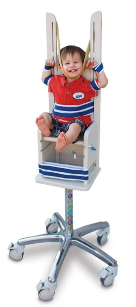
Pigg O Stat Pediatric Immobilization Gold Standard Video
The Radiology Assistant Lines And Tubes In Neonates
Paediatric Chest Immobilisation Devices Wikiradiography

Chest X Ray Of A 6 Month Old Child With An Icd The Active Can Is Download Scientific Diagram

Pedia Poser For Xray Imaging
Mountain Imaging Pigg O Stat
Ce4rt Radiographic Positioning Of The Chest For X Ray Techs

Neonatal Chest Radiograph In The Exam Setting Radiology Reference Article Radiopaedia Org

Pedia Poser For Xray Imaging
The Neonatal And Paediatric Chest Radiology Key
The Neonatal And Paediatric Chest Radiology Key
X Ray Image Classification The Easy Way By Amanda Woo Towards Data Science
Ce4rt Guide For X Ray Techs To Immobilize Pediatrict Patients
Pedia Poser For Xray Imaging
Paediatric Chest Immobilisation Devices Wikiradiography